Ventricular Septal Defects
Overview and Facts about Ventricular Septal Defects
A ventricular septal defect (VSD) is a heart condition in which a hole in the heart’s wall (the septum) separates the two lower chambers of the heart. A VSD is a common congenital heart defect (meaning it is present at birth). The hole can be in different locations in the ventricular septum, it can be different sizes, and there can also be more than one hole.
In normal fetal development, the wall between the chambers closes before birth.
Signs and Symptoms of Ventricular Septal Defects
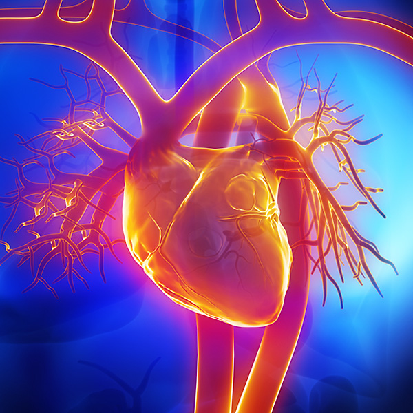
The heart has four chambers: two upper chambers known as the right atrium and left atrium, and two lower chambers called the left and right ventricles.
Normally, with each heartbeat, the right atrium receives oxygen-poor blood from the body where it then flows to the right ventricle. The right ventricle pumps the blood to the lungs to receive oxygen. Next, the left atrium receives oxygen-rich blood from the lungs and pumps it to the left ventricle. Finally, the left ventricle pumps the oxygen-rich blood to the body.
When there is a hole in the wall, oxygen-rich blood passes from the left ventricle through the opening in the septum and then mixes with oxygen-poor blood in the right ventricle. Because of this inefficient blood flow, the heart is forced to work harder.
A small ventricular septal defect rarely causes symptoms. A physician usually discovers a hole during a routine physical exam when a murmur (a swishing sound heard between heartbeats) is heard through a stethoscope.
Symptoms of larger VSDs in babies include:
- Difficulty eating
- Difficulty gaining weight
- Fast or labored breathing
- Easily tiring
When there is a large VSD, inefficient blood flow causes the heart to pump harder. Over time, the heart may enlarge and high blood pressure may develop in the arteries of the lungs (pulmonary hypertension). Symptoms may include:
- Shortness of breath and sweating
- Heart rhythm problems
- Significant fatigue
- Leg and ankle swelling (edema)
Causes and Risk Factors of Ventricular Septal Defects
This heart condition may be caused by genetic factors, problems with the heart’s development in utero, and for unknown reasons. A ventricular defect may also occur with genetic disorders such as Down syndrome.
Tests and Diagnosis of Ventricular Septal Defects
Ventricular septal defects are commonly detected during a routine physical exam; the first sign of a defect is when a murmur is heard through a stethoscope. Other tests performed may include:
- Chest X-ray
- Electrocardiogram: to evaluate the heart’s electrical system
- Transthoracic Echocardiogram with Doppler imaging: ultrasound imaging to show the flow of blood through the heart's chambers and valves.
Treatment and Care for Ventricular Septal Defects
Treatment for VSD depends on the size of the hole and severity of symptoms. Small holes with no symptoms do not require treatment; in many cases, they close on their own. In medium and large VSDs, medications may be prescribed to help the heart function better and to reduce symptoms (such as edema and heart rhythm problems).
When surgery is required, a surgical patch or stitches are used to close the hole.

Request an Appointment
Loyola Medicine heart and vascular specialists have the experience and technology to treat the most difficult cardiac and vascular conditions. Schedule an appointment today.
Schedule a Telehealth Appointment
