Cardiovascular MRI
Magnetic Imaging Test for Detailed Images of the Heart and Blood Vessels
Cardiac magnetic resonance imaging (MRI), commonly called CMR, is one of the many imaging tests performed by the highly skilled cardiologists and radiologists at Loyola Medicine.
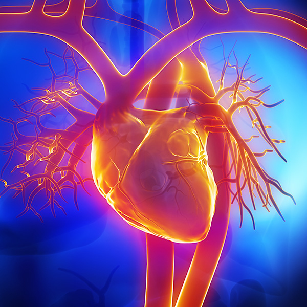
Used as a means of viewing the structure and function of your heart, a cardiac MRI is a diagnostic procedure that uses a combination of a large magnet, radio waves and a computer to take clear, detailed images of your heart and blood vessels.
Highly skilled imaging specialists are specially trained to use cardiac MRI for any of the following diagnostic purposes:
- Determine the cause of your heart failure
- Determine the extent of heart damage caused by heart attack
- Diagnose coronary heart disease
- Diagnose pericarditis
- Heart MRI in patients with pacemakers or defibrillators
- Identify heart tumor
- Monitor congenital heart disease (CHD)
- Plan an ablation procedure
- Treat irregular heart beat
Loyola is one of only a few medical centers in the state to provide you advanced cardiac MRI testing for the diagnosis and treatment of a vast array of cardiac conditions. In addition, Loyola is the only hospital in Illinois utilizing Merisight technology, in conjunction with our CMR capabilities, to provide state-of-the-art analysis for patients with atrial fibrillation.
Why Choose Loyola for Cardiac MRI?
Loyola is one of the few educational institutions in the country offering fellowships in cardiovascular imaging. All Loyola imaging specialists are fellowship-trained. Many of our physicians are involved in leading-edge research, such as the study of MRIs of patients with arrhythmias, heart failure, pacemakers and defibrillators. Several of our faculty serve on the boards of leading scientific journals.
What Should I Expect During a Cardiac MRI?
Cardiac MRI is a non-invasive procedure that creates both still and moving images of your heart and blood vessels. If your doctor requests that you receive a cardiac MRI, you will be asked to lie on a table that slides into a large scanner, which is open on both ends.
The machine uses magnetic force to align the hydrogen atoms in your body. A radiofrequency pulse is sent through you, affecting the alignment of your atoms. The signal that is generated is picked up by a scanner and interpreted by a computer to form still or moving images.
In some cases, you may receive a contrast agent, which travels to your heart and highlights its structure and blood vessels on the MRI pictures. Using contrast agent provides detailed images that can help assess the damaged areas of your heart from heart attack.
An MRI usually takes 45 to 60 minutes.
What are the Risks of a Cardiac MRI?
As a non-invasive procedure, cardiac MRI presents very few risks and complications. Because of the powerful magnet, you must be careful with metal objects in the room that has the MRI scanner. In addition, some people may have a reaction to the contrast agent if it is used.

