
Cardiac CT (Computed Tomography)
Detailed Image Scans to Detect Heart Function and Structure
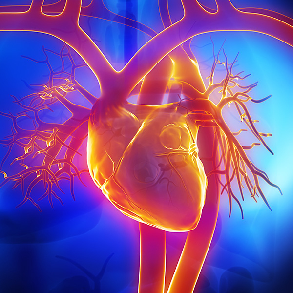
Computed tomography, commonly called a CT scan, is one of the many imaging tests performed by the highly skilled radiologists at Loyola Medicine. Used as a means of viewing the structure and function of your heart, a CT scan takes clear, detailed images of your heart structure.
A cardiac CT scan is a non-invasive procedure that takes X-ray images of your heart anatomy, coronary circulation, aorta, pulmonary veins and arteries. The machine takes a picture of each part of your heart and utilizes a computer to compile the pictures into a complete 3D image. The most common types of CT scans include:
- Calcium-score screening
- Coronary CT angiography, which utilizes a contrast agent to highlight your arteries
- Cardiac CT for evaluation, heart structure, function and valves
- CT angiography of the aorta, carotid, renal and peripheral arteries
A CT scan is generally used to diagnose and evaluate the following conditions:
- Aortic aneurysm
- Aortic dissection
- Calcium buildup
- Coronary heart disease
- Heart function problems
- Heart valve problems
- Pericardial disease
- Pulmonary embolism
- Peripheral vascular atherosclerosis
Why Choose Loyola for a Cardiac CT Scan?
Loyola is one of the few educational institutions in the country offering fellowships in cardiovascular imaging. All Loyola cardiologists and radiologists are fellowship-trained. Many of our physicians are involved in leading-edge research, such as the study of cardiac CT in evaluation of chest pain and valvular heart disease. Several of our faculty serve on the boards of leading scientific journals.
What Happens During a CT Scan?
A cardiac CT scan is a painless procedure requiring minimal preparation. If your doctor plans to use IV contrast during your procedure, you may be asked to limit your food intake for four to six hours prior to your scan.
During the procedure, you will be asked to lie down on a table and electrodes will be connected to your chest, enabling the doctors to monitor your heart. The table you are on will be moved into the machine and the scanner will begin taking pictures by moving around your body. The scanner will not touch you.
The procedure length is generally 10 to 15 minutes in duration and you are able to resume normal activity following the scan.
What are the Risks of a CT Scan?
Your doctor will discuss with you the risks and benefits of getting a cardiac CT scan, but generally the risks are minimal. Due to the use of an X-ray machine, you will be exposed to radiation, but the amount used is considered minimal—and the benefits of CT scans for certain conditions outweigh the risks. In some instances, you may develop an adverse reaction to the contrast agent used during the scan.
Request an Appointment
Loyola Medicine heart and vascular specialists have the experience and technology to treat the most difficult cardiac and vascular conditions. Schedule an appointment today.
Schedule a Telehealth Appointment