
Cornea and External Disease
Overview and Facts about Corneal and External Eye Disease
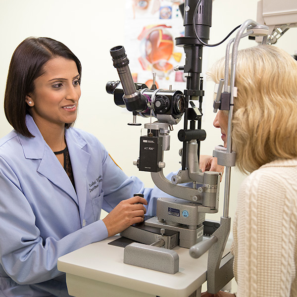
Two fellowship trained corneal surgeons in the department of ophthalmology are committed to providing the very best medical and surgical care to patients who present with a wide variety of corneal, conjunctival and refractive eye diseases through integrated, patient-centered care teams.
We perform the latest diagnostic testing and surgical techniques, including endothelial keratoplasty (EK), lamellar keratoplasty (LKP), penetrating keratoplasty (PKP), LASIK and photorefractive keratectomy (PRK) as well as ocular surface reconstruction techniques including mucous membrane and amniotic membrane grafting. We also manage patients referred for complex cataract (and cataract complications) and other anterior segment reconstruction procedures.
Our Loyola Ocular Surface Disease Center (OSDC) is dedicated to provide comprehensive, multidisciplinary and structured care for patients with a variety of ocular surface diseases, including corneal stem cell deficiency, ocular graft versus host disease (oGVHD), Stevens Johnson syndrome (SJS), thermal and chemical injury and severe tear dysfunction syndrome (TDS).
Comprehensive evaluation includes an ocular surface disease index (OSDI) assessment, a complete and thorough eye exam and the latest technology in the diagnosis of these conditions, including meibography, topography, tomography, Schirmer testing, tear break up time and tear film inflammation testing (InflammaDry®).
A detailed and personalized management plan is provided with helpful online resources and readable handouts. This plan often includes complex contact lens management, serum tears, amniotic membrane and limbal stem cell transplantation.
Research about Cornea and External Disease
The Cornea and Ocular Surface Disease Research Program is part of the larger Richard A. Perritt Eye Research and Education Collaborative, which focuses on four research areas: education technology, corneal and ocular surface diseases, glaucoma and neurodegenerative retinal disease, and bioinformatics and patient care outcome research.
Our cornea research faculty are working on numerous projects investigating mechanisms and treatment of:
- Inflammatory Dry Eye Disease (DED)
- Stevens Johnson Syndrome (SJS)
- Keratoconus (KCN)
- Ocular Graft versus Host Disease (oGVHD)
- Ocular manifestations of obstructive sleep apnea (OSA)
- Endothelial dysfunction and keratoplasty (EK)
Learn more about Loyola's cornea and external disease research
What Conditions Does Loyola Treat?
Loyola’s eye specialists offer a full range of services to treat corneal and external eye diseases and conditions, including:
- Routine cataract surgery
- Complex cataract surgery including patients with dry eye or following LASIK surgery
Conjunctivitis involves inflammation or swelling of the conjunctiva, the tissue overlying the white part of the eye and inner surface of the eyelids. Causes include bacteria, viruses, allergy, and chemical exposure. Symptoms may include redness, discharge, tearing, swelling, light sensitivity, itching and burning. Some types of conjunctivitis are contagious and can spread quickly. Treatment depends on accurate diagnosis of the cause of conjunctivitis.
These include infection, allergy, dry eye, corneal abrasions, and more complex problems including corneal ulcers and corneal stem cell deficiency.
There is a wide range of ocular trauma including mechanical, thermal and chemical injury. The trauma can be mild, moderate or severe and can effect all parts of the eye leading to mild, moderate or severe vision loss.
Corneal ulcer is an infection of the cornea, the outer clear layer of the eye in front of the iris. Most commonly, corneal ulcers are due to bacterial infection, but they can also result from infections due to fungi and parasites. Contact lens wearers are at increased risk of corneal ulcers, especially those who sleep or swim with their lenses in.
Dry eye disease (DED) is caused by a chronic lack of sufficient lubrication and moisture on the surface of the eye. This can result from inadequate quality or quantity of tear production as well as an increase in evaporation of ears. Consequences of DED range from subtle but constant eye irritation, blurred vision, and eye redness to significant inflammation and even scarring of the cornea.
Fuchs corneal dystrophy is a disease of the cornea. It involves dysfunction of the endothelial cells, the inner lining of the cornea responsible for pumping fluid out of the cornea and keeping the cornea thin and clear. When the endothelial cells lose function, the cornea becomes swollen, causing blurry vision. Early on, this results in morning blurriness that then improves during the day. Advanced disease leads to chronic loss of vision and may require partial thickness transplantation of the inner lining of the cornea (DSEK).
Keratoconus is a common corneal disorder where the central cornea undergoes progressive thinning and steepening causing irregular astigmatism and blurred vision. Etiology is unknown. However, it is associated with atopy, Down’s Syndrome, Leber’s congenital amaurosis, and Ehler’s Danlos/connective disorders. Risk factors include eye rubbing, obstructive sleep apnea, and floppy eyelid syndrome. Treatments include contact lenses and a variety of surgical and laser procedures.
Floppy eyelid syndrome (FES) is an under-diagnosed condition of distensible eyelids due to a decrease in elastin. It effects 20% of the US population and can be associated with dry eye irritation. Floppy Eyelid Syndrome is associated with obesity, keratoconus and obstructive sleep apnea (OSA). OSA results from intermittent upper airway blockage resulting in chronic systemic inflammation. This systemic inflammation can lead to a variety of eye diseases including stroke in the retina, optic nerve and low tension glaucoma. It is also associated with other systemic diseases such as stroke, cardiac arrhythmias, and sudden cardiac death.
- Meibomian gland dysfunction (MGD) describes the blockage and or inflammation of the Meibomian glands in the eyelids so they don't secrete the normal amount and/or quality of oil into the tear film. This may result in alteration of the tear film, symptoms of eye irritation, clinically apparent inflammation, and ocular surface disease. The oily layer produced by the Meibomian glands normally slows down tear evaporation so any abnormality in these glands can lead to dry eye. MGD is actually the leading cause of dry eye disease (DED).
- Blepharitis is a chronic inflammation of the eyelids, which causes redness and matting of the eyelids. It has a variety of causes, ranging from allergy and infection to irritation.
Ocular Graft Versus Host Disease (GVHD) occurs in patients who have undergone allogenic hematological stem cell transplantation. It can occur in patients who have acute or chronic GVHD, though it is more common in patients with the chronic form. Symptoms are not unique to this disease and can include dry eye, foreign body sensation, redness and irritation. It can also cause tearing , light sensitivity, blurred vision and severe pain. Children with a history of bone marrow transplantation can also develop ocular symptoms but do not complain of the same symptoms as adults. Often children are noted to have excessive eye rubbing and light sensitivity as their predominant symptoms.
Scleritis is painful inflammation of the sclera, the white part of the eye. In many cases, it is due to an underlying autoimmune condition. There can be both anterior and posterior scleritis. Treatment involves system anti-inflammatory medications, including prednisone and immune-modulating agents.
Stevens-Johnson syndrome (SJS) and its more severe variant, toxic epidermal necrolysis (TEN) are a disease continuum of potentially life-threatening, severe allergic drug reactions resulting in the destruction of the skin, mucous membranes and ocular surface.
Tearing (“epiphora”) can be associated with a variety of changes in the ocular surface, including blocked tear drainage ducts and sometimes evaporative dry eye, which gets worse in the wind or other environmental insults.
How is Cornea and External Disease Diagnosed?
High resolution imaging of the cornea and front part of the eye
Indications: Anterior segment OCT can be used for multiple reasons, including evaluation of corneas before and after corneal transplantation/refractive surgery, evaluation of corneal or conjunctival lesions, and evaluation of pathology of the anterior chamber and angle.
Procedure: The patient is positioned in a machine similar to a slit lamp, and a quick photo is taken of each eye separately.
High resolution imaging of the corneal surface
Indications: Corneal topography may be done on patients with keratoconus or other corneal ectasias, as well as patients planning to undergo cataract surgery or refractive surgery.
Procedure: After the patient is positioned correctly in the machine, a quick photo is taken of each cornea.
High resolution imaging of the corneal thickness
Indications: Corneal tomography creates a 3D model of the patient’s cornea which is useful in patients with keratoconus or other corneal ectasias, as well as patients planning to undergo cataract surgery or refractive surgery.
Procedure: After the patient is positioned correctly in the machine, a quick photo is taken of each cornea.
Corneal ultrasound thickness measurement
Indications: Corneal thickness can be measured to evaluate patients with Fuchs’ dystrophy, corneal transplants, other causes of corneal edema, and glaucoma patients.
Procedure: After an anesthetic eye drop is placed in the eye, an ultrasound probe is used to measure the corneal thickness.
Immunohistopathological testing
Indications: Immunohistopathological testing can help in the diagnosis of ocular surface conditions such as ocular cicatricial pemphigoid, conjunctival nevus, primary acquired melanosis, melanoma, and squamous cell carcinoma.
Procedure: Either in the minor procedure room or main operating room, samples are taken from the ocular surface after anesthetic drops are placed in the eye. The samples are then sent to the laboratory for evaluation.
Imaging of the lipid (oily) part of the tear film and the eyelid Meibomian glands
Indications: Meibography can be very useful in the evaluation of patients with dry eyes or symptoms of burning, irritation, and tearing.
Procedure: After the patient is positioned correctly in the machine, several photos and videos are taken of the eyes.
Corneal cultures for bacterial, viral, and parasitic (amoebic) organisms
Indications: Microbiological testing is critical in the diagnosis and evaluation of patients with corneal ulcers and other ocular surface infections.
Procedure: After anesthetic eye drops are placed, a swab is used to take a sample from the ulcer and then placed on multiple culture plates. These plates are then sent to the laboratory for evaluation.
High resolution photos of the front part of the eye
Indications: Slit lamp photos can be useful to document and follow conjunctival and corneal lesions such as pigmented lesions, pterygia, corneal ulcers, and epithelial ingrowth.
Procedure: The patient is positioned in a slit lamp machine with a camera attached, and several photos are taken.
Quantitative imaging of the number of cells on the inner lining of the cornea
Indications: Specular microscopy is helpful in the evaluation of patients with Fuchs’ dystrophy and other corneal endothelial disorders, as well as monitoring endothelial cell count before and after surgery including cataract surgery and corneal transplantation.
Procedure: The patient is positioned in the specular microscopy machine, and photos are taken of each eye.
InflammaDry – levels of matrix metalloproteinase-9 levels (MMP-9)
Indications: This test can be performed to evaluate level of inflammation in eyes with dryness or other ocular surface diseases.
Procedure: The InflammaDry stick is touched to the surface of the eye and then placed into a cartridge, very similar to a pregnancy test.
LipiView II Ocular Surface Interferometer
The LipiView® II Ocular Surface Interferometer is a device which measures 3 important tear functions.
- Lipid Layer Thickness (LLT)
- The thickness (accurate to ±10 nm) of the front oily layer of the tear film. This layer reduces tear film evaporation associated with “dry eye” symptoms. The results are presented in an easy to understand color-coded map.
- Dynamic Meibomian Gland Imaging
- Provides high-definition images of the anatomy and structure of the meibomian (oil) glands in each lower eyelid
- Blink Dynamics
- Analyzes blink patterns and detects partial blinks. A graphical representation shows fluctuations in lipid layer thickness measurements between each blink.
Schirmer test, tear break up time, tear film meniscus
Indications: These tests are useful in diagnosing and monitoring patients with dry eye symptoms. Schirmer test is used to measure tear production. Tear break up time looks at tear film instability. Tear film meniscus can help quantitate the amount of tears in the eye.
Procedure: After topical anesthetic drop is placed in each eye, a small strip of paper is placed in the corner of each eye. After 3 minutes, the strips are removed and the tear production is measured. Tear break up time is measured by placing a fluorescein drop in each in eye, having the patient blink, and then counting the number of seconds until the first dry spot is noted on the cornea. Tear film meniscus is measured by looking at the height of the tear film using the slit lamp.
How is Cornea and External Disease Treated?
We offer surgical treatment for a variety of complex anterior segment conditions using the latest treatment techniques, including:
- Amniotic membrane grafts
- Onlay (biological patch dressing for acute Stevens Johnson Syndrome and neurotrophic keratitis)
- Inlay (for corneal thinning disorders)
- ProKera (for inflammatory corneal disease)
- Autologous serum tears (for dry eye and inflammatory corneal disease)
- Epithelial debridement (for recurrent corneal erosion syndrome and basement membrane corneal dystrophy)
- Corneal limbal stem cell transplantation
- Penetrating Keratoplasty (full thickness)
- Lamellar keratoplasty (partial thickness)
- Descemet’s stripping endothelial keratoplasty (DSEK) - transplantation of the inner most layer of the cornea
- Descemet’s membrane endothelial keratoplasty (DMEK)
- Refractive Surgery
- LASIK
- Photorefractive keratectomy (PRK)
- Phototherapeutic keratectomy (PTK)
- Pterygium surgery (for primary, recurrent, and double pterygia)
- Fornix reconstruction
- Buccal mucosal transplantation (for eyelid margin scarring)
- Symblepharon repair
- Amniotic membrane grafts
- Repair of exposed scleral buckles
- Conjunctivochalasis repair
- Cataract surgery
- YAG laser capsulotomy
- Insertion of capsular tension rings (CTR)
- Pediatric cataract surgery
- Complex cataract surgery
- Pseudoexfoliation syndrome
- Post LASIK cataract surgery
- Post RK cataract surgery
- Cataract in patients with Marfan’s Syndrome
- Post-traumatic cataract
- Zonular dehiscence
- Secondary intraocular lens (IOL) implantation
- Anterior chamber placement
- Iris fixation of IOL
- Scleral fixation of IOL
- Scleromalacia repair
- Fascia lata patch grafts
- Scleral patch grafts
- Lamellar corneal patch grafts
- Tutoplast® grafts
- Amniotic membrane grafts
Request an Appointment
Whether you are seeking routine eye care or have a specific vision issue, our team treats a wide range of eye diseases and conditions, including cataracts, glaucoma, macular degeneration and strabismus. Schedule an appointment today.
Schedule a Telehealth Appointment