
Retina/Diabetic Eye Care
What Conditions Does Loyola Treat?
Diabetic macular edema (DME) is an ophthalmological condition that affects your eyesight and is a complication of a condition called diabetic retinopathy. Individuals with diabetes are often at risk of developing diabetic retinopathy, which can occur when your blood sugar levels are poorly controlled.
Over time, diabetic retinopathy damages the blood vessels in your eyes. The blood vessels may swell or leak, causing fluid and pressure to build up in the eye. DME develops when fluid seeps into the part of the retina known as the macula.
The retina is the part of your eye responsible for focusing your vision. When the retina swells, your vision may become blurry or distorted. DME affects up to 10 percent of people with diabetes. The condition is particularly common among individuals with Type 1 diabetes. If you are diabetic, the best way to protect your eyesight from diabetic retinopathy is to detect the disease early on. Even if you don’t have any symptoms, you should have eye examinations once a year. Once you are diagnosed with diabetic retinopathy, your follow-up exams may be more frequent.
Epiretinal membrane (ERM), sometimes referred to as a macular pucker, is fibrous tissue that develops on the surface of the eye’s retina in the macular area. Depending on the severity of the condition, ERM can cause disturbances in vision, notably central vision, or no symptoms at all. If you notice changes in your vision, it’s important to consult your physician.
A macular hole is a small hole in the macula, a part of the eye in the middle of the retina. The macula is made up of light-sensitive tissue that helps the eye focus; a macular hole results in blurry or distorted vision. There are three different stages of macular holes. Stage I, also known as foveal detachments, are the least serious. Without treatment, about 50% of Stage I macular holes progress to Stage II, or partial-thickness holes. Without treatment, 70% of Stage II macular holes become Stage III and Stage IV or full-thickness holes.
Retinal detachments affect about one in 10,000 people. They typically happen to patients who are nearsighted, have a family history of retinal detachments and have had cataract surgery.
The retina is a thin layer of nerve tissue that lines the inside of the back of the eye. Its role is to help the eye communicate with the brain, which occurs when light-sensitive photoreceptors known as rods and cones convert light into electrical impulses that are carried to the brain through the optic nerve. The brain then translates these electrical signals into images.
A retinal tear occurs when the vitreous (gel-like material that fills the inner eye and is attached to the retina) forcibly pulls away from the retina, causing a tear in one or more places.
Retinitis pigmentosa is a series of ophthalmological conditions that break down the retina. Because the retina is made up of light-sensitive tissue that helps you see, one of the primary symptoms of retinitis pigmentosa is loss of sight. Retinitis pigmentosa is an inherited disease, passed down from a parent to a child. Overall, it’s a relatively rare disorder, affecting 1 out of every 4,000 people.
Central serous chorioretinopathy (CSC) is a condition in which fluid builds up behind the retina. This fluid comes from the choroid, a layer of tissue under the retina, and leaks through the retinal pigment epithelial (RPE) into the retinal space. CSC causes blurred vision in one or both eyes and may lead to temporary or permanent vision loss. The cause of this condition is unknown and it typically affects men in middle age.
How Are Retina/Diabetic Eye Conditions Diagnosed?
To diagnose issues with the retina or eye issues related to diabetes, your physician may use a variety of different tests, including:
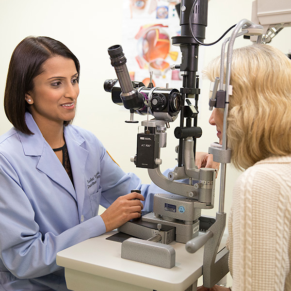
A dilated eye examination allows your ophthalmologist to see the back of your eye, including the retina. During this exam, your physician uses an ophthalmoscope (an instrument with a bright light and special lens) to examine the back of the eye for signs of abnormal blood vessels, retinal detachment and bleeding.
A visual field test determines any changes to your peripheral vision.
This test involves taking pictures of your eyes before and after a special dye called fluorescein is injected into your arm. Your ophthalmologist first dilates your eyes, and then takes pictures of the inside of your eyes. Your arm vein is then injected with the fluorescein dye and more pictures are taken as the dye circulates through your eyes’ blood vessels. Your ophthalmologist can use the images to pinpoint blood vessels that are closed, broken down or leaking fluid.
This test is used to diagnose a retinal tear. It is also used if there is any bleeding in the eye, which makes it difficult to see the retina in a routine eye exam.
An optical coherence tomography (OCT) exam helps your ophthalmologist determine the thickness of your retina, which will reveal whether fluid has leaked into retinal tissue. OCT exams can also be used to monitor how treatment is working.
How Are Retina/Diabetic Eye Conditions Treated?
Treatment for retina conditions, including diabetic retinopathy, includes active monitoring, medication and surgical options.
Medication injections
Medications called vascular endothelial growth factor (VEGF) inhibitors may help stop growth of new blood vessels. They work by blocking growth signals the body sends to create new blood vessels.
Loyola’s skilled ophthalmic surgeons are experienced in the following surgical techniques:
In photocoagulation, the surgeon directs a laser into the eye through the pupil to reduce abnormal blood vessel growth and help attach the retina to the back of the eye. This treatment is used for diabetic retinopathy, bleeding into the vitreous and retinal detachments.
After the eye is numbed with a local anesthetic, the surgeon applies a freezing probe over the retinal tear, which causes a scar that helps secure the retina to the eye wall.
For patients with extensive detachment, scleral buckling may be the preferred treatment. In this procedure, your physician will relieve the pressure caused by the detached retina using a piece of silicone sponge, rubber or plastic to hold the eye in place.
This is a laser treatment used to shrink abnormal blood vessels in the eye. In this procedure, the areas of the retina away from the macula are treated with scattered laser burns, which cause the abnormal new blood vessels to shrink and scar.
Your doctor may recommend pneumatic retinopexy to reattach or repair your retina. In this procedure, your doctor will inject a bubble of air or gas into the center of the eye, which will surround and seal the area of concern. This gas bubble will stop the flow of fluid behind the retina and dissolve on its own.
Your doctor may recommend vitrectomy, which removes the gel-like vitreous tissue from the center of the eye, and injects air, gas or liquid to reattach the retina. This treatment is used for retinal tears/detachment, ERM, macular holes and diabetic retinopathy.
Request an Appointment
Whether you are seeking routine eye care or have a specific vision issue, our team treats a wide range of eye diseases and conditions, including cataracts, glaucoma, macular degeneration and strabismus. Schedule an appointment today.
Schedule a Telehealth Appointment