
Retina and Vitreous Diseases
What Conditions Does Loyola Treat?
Central serous chorioretinopathy (CSC) is a condition in which fluid builds up behind the retina. This fluid comes from the choroid, a layer of tissue under the retina, and leaks through the retinal pigment epithelial (RPE) into the retinal space.
CSC causes blurred vision in one or both eyes and may lead to temporary or permanent vision loss. The cause of this condition is unknown and it typically affects men in middle age.
Diabetic retinopathy is a complication of diabetes that affects the eyes. It can develop in anyone with type 1 or type 2 diabetes, causing damage to the blood vessels in the retina.
Initially, this condition may cause no symptoms and only slight vision problems. If it is not treated promptly, it can result in other ophthalmology conditions, including blindness.
Macular degeneration is an ophthalmological condition that results when a part of your retina (known as the macula) deteriorates. The retina, located at the back of the eye, helps focus your vision. In most cases, aging is the cause of macular degeneration. Age-related macular degeneration is the most common cause of vision loss in older adults.
There are two types of macular degeneration: the dry-type that causes a gradual loss of vision and the wet-type that causes a sudden loss of vision.
The retina is a slim, light-sensitive layer of tissue that lines the inside of your eye and sends messages to your brain through the optic nerve, allowing you to see. When the retina detaches or moves away from the eye, it is an emergency situation that calls for immediate medical attention in order to save or restore your vision. There are three types of retinal detachment:
- Exudative retinal detachment occurs after an injury or because of diseases that cause inflammation.
- Rhegmatogenous retinal detachment, the most common of the three types of detachment, occurs when a tear in the retina allows fluid to accumulate and interfere with retinal nourishment.
- Tractional retinal detachment occurs when scar tissue or other damage causes the retina to contract away from the retinal pigment epithelium (RPE).
Vitreous is a gel-like substance in your eye that helps it maintain its round shape. As you age, the fibers within the vitreous that are attached to your retina shrink. When these fibers shrink so much that they break, the vitreous can separate from the retina, causing vitreous detachment.
Symptoms of vitreous detachment include seeing “floaters” and/or flashes. Floaters are strands from the vitreous that cast shadows in your field of vision, and flashes of light may accompany floaters in your peripheral vision.
Vitreous detachment can be diagnosed with a dilated eye exam, which will help your physician determine if treatment is necessary. This condition is not sight-threatening and in most cases requires no treatment. If, however, vitreous detachment has caused a macular hole, retinal tear or retinal detachment, prompt treatment is required to prevent vision loss.
How are Retina and Vitreous Diseases Treated?
Indications: This test helps your doctor visually check any abnormal blood vessels in your eyes. An angiography test requires your eyes to be fully dilated.
Procedure: During this procedure, a special dye is injected into your veins. As the dye passes through the blood vessels in your eyes, your doctor takes photographs to help reveal the existence of abnormal blood vessels in your eyes.
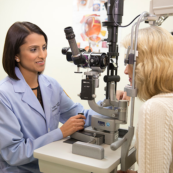
Indications: The diagnosis of retina and vitreous disease is made with a full-dilated eye examination.
Procedure: A dilated eye examination allows your ophthalmologist to see the back of your eye, including the retina. During this exam, your physician uses an ophthalmoscope (an instrument with a bright light and special lens) to examine the back of the eye for signs of abnormal blood vessels, retinal detachment and bleeding.
Indications: An optical coherence tomography (OCT) exam helps your ophthalmologist see each of your retina’s layers to determine the thickness of your retina, which will reveal whether fluid has leaked into retinal tissue. OCT exams can also be used to monitor how treatment is working for many eye conditions, including macular degeneration, glaucoma, central serous retinopathy, diabetic retinopathy and macular edema.
Procedure: During an OCT exam, your ophthalmologist will administer eye drops to dilate your pupils. You will sit in front of an OCT machine and the equipment scans your eye with infrared light for 5-10 minutes without touching it.
Indications: Vision tests can help determine whether your eyesight is blurry or distorted.
Procedure: Your doctor will position you in front of a grid chart and ask you to alternate between closing each eye, asking you to indicate when the lines look wavy or distorted. They will mark any defects that you indicate and assess if your answers have changed since your last visit.
How Are Retina and Vitreous Diseases Treated?
Treatment for retina conditions includes active monitoring, medication and surgical options.
In laser surgery for retina and vitreous disease, the surgeon uses a laser to heat small points on the retina to attach the retina to the back of the eye. This treatment is used mainly for retinal tears and detachments. Laser surgery can also be used to treat diabetic retinopathy.
Intravitreal injections may be used to treat retina and vitreous diseases and preserve your vision. During this procedure, the ophthalmologist injects medication into the vitreous after an anesthetic has been administered.
This procedure is often used for macular degeneration and diabetic retinopathy.
Request an Appointment
Whether you are seeking routine eye care or have a specific vision issue, our team treats a wide range of eye diseases and conditions, including cataracts, glaucoma, macular degeneration and strabismus. Schedule an appointment today.
Schedule a Telehealth Appointment