
Glaucoma
What Glaucoma Conditions Does Loyola Treat?
Acute angle closure glaucoma occurs when there is a sudden rise in intraocular pressure, the pressure inside the eye. This is caused by a buildup of fluid in the eye. Normally, fluid drains out of the eye through a channel called the angle that runs between the cornea and the iris. In cases of acute angle closure glaucoma, the iris presses against the angle, closing it off and preventing fluid from being released.
Chronic angle closure glaucoma develops slowly over time as the iris, or colored part of the eye, blocks the eye’s drainage angle. Once it becomes fully blocked, the pressure in your eye increases, leading to a range of symptoms and, sometimes, permanent eye damage.
Primary open angle glaucoma is the most common of the glaucoma conditions. As the condition progresses, your eye pressure increases; however, your cornea doesn’t swell and you remain symptom free. This often results in the condition going undetected, until permanent damage occurs. Once the optic nerve becomes damaged, you may notice blind spots or loss of vision, especially in your peripheral or side vision.
Pseudoexfoliation glaucoma is an eye disease that is characterized by the presence of an exfoliative material in the anterior portion of the eye. Over time, this exfoliative material, which is largely comprised of abnormal and thin fibers that overlap each other, builds up in the anterior portion of the eye, and causes eye pressure to rise leading to optic nerve damage. Damage to the optic nerve results in impaired or loss of vision, or glaucoma.
Pigmentary glaucoma is an eye condition that occurs when the layer of the eye containing pigment, the iris, rubs against the lens, causing the release of pigment particles. These particles accumulate in the eye, preventing the fluid produced by the eye from draining. Fluid buildup can cause increased eye pressure and the development of glaucoma conditions. Eventually, damage to the optic nerve, the structure responsible for sending visual stimuli to your brain, leads to vision loss.
Angle recession glaucoma is a condition that develops from blunt trauma to the eye. Injury can cause the fluid inside your eye to be displaced, which may cause damage that includes torn eye tissues. As a result, scarring and degenerative changes can occur, which blocks the channel that drains the fluids produced by your eye. Fluid buildup can further cause elevated intraocular pressure (pressure inside the eye), leading to angle recession glaucoma.
Neovascular glaucoma is a condition that affects your eyes’ blood vessels and is frequently associated with diabetes. In neovascular glaucoma, the vessels that supply oxygen-rich blood to your eyes are constricted, thereby cutting off the oxygen necessary to function. Consequently, your body will signal new blood vessels to grow. This process eventually causes new growth to plug the channel that drains the fluids produced by your eye. Blockage can cause fluids to build up and create pressure on the optic nerve; damage to the nerve will eventually lead to vision loss.
Congenital glaucoma occurs when high fluid pressure in the eye puts too much strain on the optical nerve, damaging it. Normally, this fluid is drained away from the optical nerve, but people with this disease are born with a problem that affects the eye’s drainage channel. Because this disease is present at birth, this problem is either due to an issue with development or inherited malformation.
How Is Glaucoma Diagnosed?
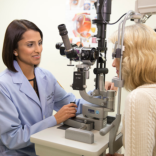
Indications: A slit lamp microscope is used to determine whether or not there are changes in the optic nerve’s size and shape.
Procedure: The patient is positioned in front of the slit lamp and the ophthalmologist focuses a narrow, high-intensity beam towards the eye to examine the optic nerve.
Indications: Glaucomatous damage produces characteristic defects in the central and peripheral visual fields. Vision tests can be used to assess any abnormalities that may indicate the presence of glaucoma.
Procedure: The patient is asked to look straight ahead and indicate when a moving light passes into the peripheral visual field.
Indications: Normal IOP is between 12-22 mm Hg (millimeters of mercury). Elevated IOP is a major risk factor for the development of glaucoma. Optic nerve damage becomes more likely as the IOP increases.
Procedure: The patient’s eyes are numbed with eye drops and the inner pressure of the eye is measured with a device called a tonometer or a warm puff of air.
Request an Appointment
Whether you are seeking routine eye care or have a specific vision issue, our team treats a wide range of eye diseases and conditions, including cataracts, glaucoma, macular degeneration and strabismus. Schedule an appointment today.
Schedule a Telehealth Appointment